The FAST exam is a point of care ultrasound protocol indicated for the rapid assessment of trauma patients. Specifically it looks for free fluid (usually blood) in the abdomen and pericardial sac. An extended scan, eFAST, also includes intercostal views for assessment of the pleural space. There are no absolute contraindications to eFAST scanning, but it should not delay resuscitative efforts to a patient in extremis.
The scan results are used in the following algorithm.

Views
- RUQ
- LUQ
- Pelvic (transverse and logitudinal)
- Subxiphoid/subcostal
- Intercostal (right and left, 2/3rd intercostal space)
RUQ
This view is the easiest as the liver is a large acoustic window. Abdominal free fluid often collects here first, or in the pelvis. Ensure the tip of the liver is visualised as free fluid there is often missed if the collection is small.
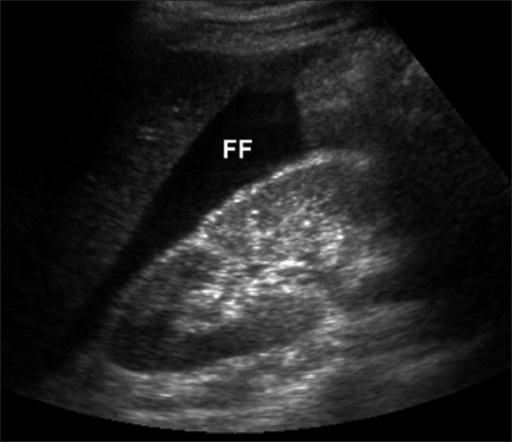
Jyothi PSLS, Kalra VB - CC BY-NC-ND 4.0, Link
LUQ
The spleen is much smaller than the liver and thus the RUQ acoustic window is small: this view is technically more difficult to obtain. Collections form between the spleen and diaphragm. The spenorenal interface is preserved by the splenorenal ligament.
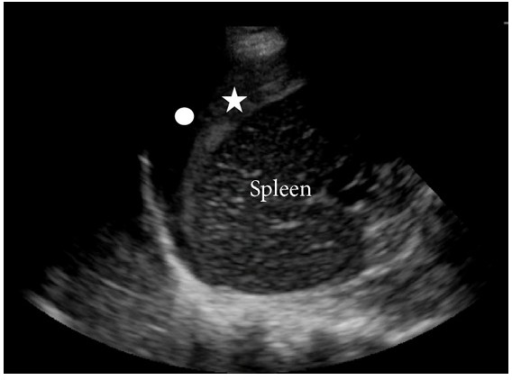
The star indicates clotted blood adherent to the spleen. Jackson H, et al. - CC BY 3.0, Link
Pelvis
A dependent collection in the pelvis will be visualised in the retrovesical pouch or the pouch of Douglas in females.
Subcostal view
The subcostal view can also be difficult to obtain. In the FAST scan, visualisation of the chambers of the heart is less important; the primary aim is assessment of the pericardial space.
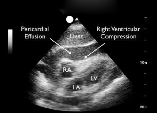
Note the obliteration of the right ventricle. Goodman A, et al. - CC-BY-SA 3.0 Link
Lung views
The opposition of pleural and aerated lung below it produces a characteristic image as the parietal and visceral pleura slide against each other, often described as “marching ants”. An M-mode spike through the intercostal window will produce another characteristic temporal image known as the “seashore sign”.
In the setting of pneumothorax, the M-mode image will become a “barcode sign”. This view is less valuable in the identification of haemothorax or other intrapleural free fluid, they are probably better looked for at the diaphragm in the RUQ and LUQ views.
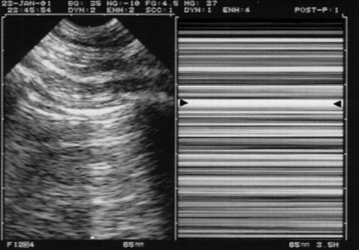
Right: M-mode, the abolition of lung sliding is visible as the “Barcode sign”. The arrows indicate the pleural signal. Lichtenstein DA - CC-BY 3.0 Link